Philips HD11 XE Ultrasound Machine
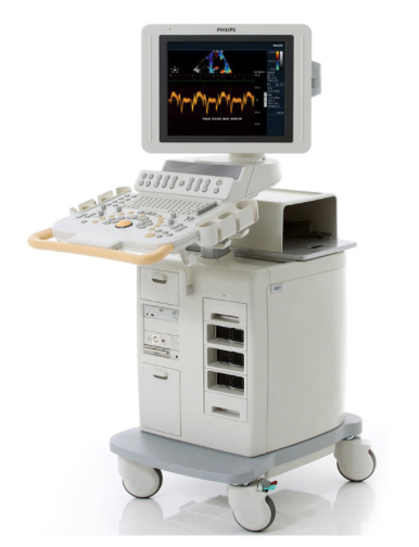
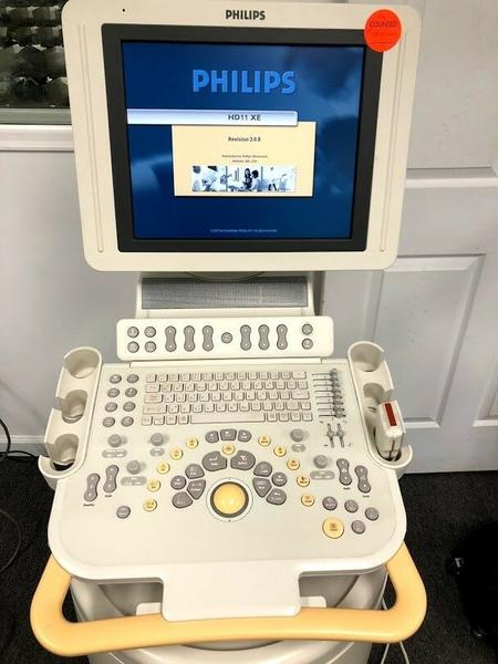
The Philips HD11xe is an extremely popular midrange ultrasound machine that dominated sales in the shared service segment for nearly a decade. The HD11xe balances price and performance perfectly while still giving access to high-end features such as 4D and TEE. The HD11xe had more transducer options than any other midrange and even more than many high-end ultrasound machines. The HD11xe is the replacement for the HD11 and adds an LCD monitor on an articulating arm and software upgrades such as Qlab. The HD11xe is still in high demand after it has gone out of production and offers an amazing value as a refurbished or remanufactured system.
The HD11xe, while specializing in cardiovascular ultrasound applications, is also capable of producing images for other general applications. This includes OB-GYN, small parts, musculoskeletal and abdominal applications.
Product Features:
- Excellent diagnostic imaging capability in all major clinical applications. A simply good and inexpensive ultrasound unit.
- Flexible Architecture-Broadband digital transducers and beamformer capture and preserve the entire bandwidth of ultrasound signals to retain the quantity and quality of tissue signatures, while advanced signal processing fully uses the diagnostic information within the broadband digital signal without sacrificing temporal resolution.
- Proven Technologies - SonoCT imaging processes up to nine lines of sight, for more diagnostic information, while XRES adaptive technology performs 350 million calculations per frame for superb resolution.
- Easy to move - Excellent mobility and portability, as well as a rotating, height-adjustable control panel and monitor for advanced ergonomics.
- Easy to use - Integrated automation tools make it easy to achieve the superb 2D, Doppler or 4D image with minimal keystrokes.
- Imaging and data management - A suite of Advanced Imaging modes deliver diagnostic power, for complete exam protocols with incredible system responsiveness. Data management capabilities allow flexible recording, archiving, editing and even exam reports with embedded images.
- 2D with Pulse Inversion Harmonic Imaging, Philip's patented method for producing pure, broadband harmonic signals for superb grayscale presentation.
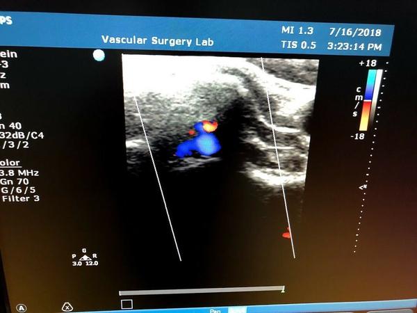
- Adaptive Color Doppler, which automatically selects the optimal Doppler or angio frequency for highly sensitive resolution, as well as Color Power Angio technology for assessing amplitude and direction of flow.
- Pulsed wave and continuous wave Doppler with Adaptive Doppler technology to boost weak signals and reduce noise, and high PRF capability for measuring higher velocities than ordinary pulsed Doppler ultrasound.
- Tissue Doppler Imaging (TDI), including Color TDI to assess direction and timing of myocardial function, and pulsed wave TDI for velocity mapping of vessel wall motion and cardiac tissue.
- Anatomical M-mode, for more accurate measurements of chambers, walls, and ejection fraction; makes it easier to keep the M-mode line perpendicular to the anatomy, even in abnormally shaped or positioned hearts.
- Q lab advanced Quantification
- 3D/4D imaging - The HD11 brings 3D/4D imaging into the mainstream by delivering it on a platform with an excellent combination of versatility and value.
- 3D fetal echo STIC - Allows for a more detailed view of fetal heart valves and wall motion, to aid in detecting anomalies during routine obstetrical exams.
- Performance features - With optional panoramic imaging, contrast imaging, stress echo, and DICOM Networking, you can design the HD11 to fit your specific clinical needs.


