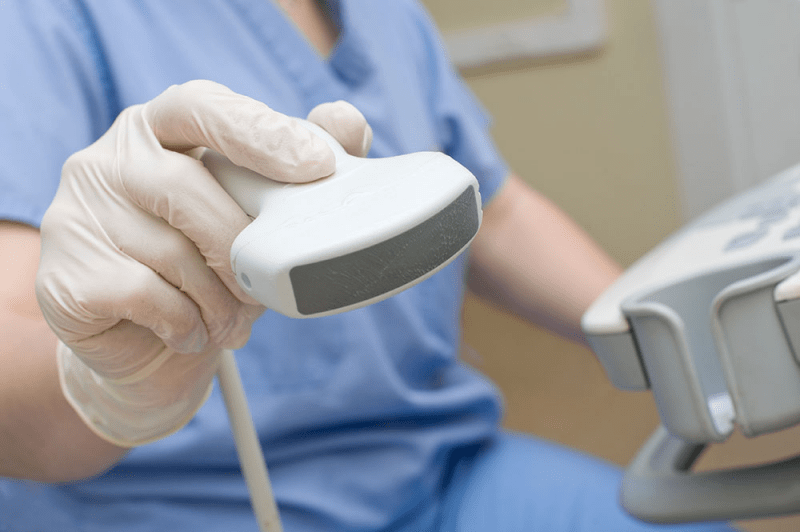
What is a Probe / Transducer?
Ultrasound probes serve as the primary tools for Ultrasound machines. Without the probes, the Ultrasound machine is incapable of producing or providing images. The ultrasound machine ultimately serves as a translator between people and the transducer. The transducer generates non-invasive sound waves that are unable to be processed by human ears. The sounds reverberate back, and the ultrasound system converts the sounds into images of the particular structure that the sound bounced off of. Without the image, sonographers and diagnosticians are unable to perform even the most basic functions of their jobs.
Now that we understand the roles that the ultrasound machine and the transducer / probe play in diagnostic imaging, respectively, we can understand why the probe is integral. Without a probe that functions properly, the images that the system translates are inherently unreliable. Attempting to diagnose a patient without accurately assessing the symptoms is dangerous – equally as dangerous as attempting to diagnose internal organs and structures without accurate images.
Maintaining A Transducer:
Transducers, extremely delicate in nature, need to be kept in top condition. Even the slightest nick or dent in the lens can pervert and damage the image quality on the screen. Simply put, using a combination of sound waves and electric waves, the probe generates voltage that translates into images on the Ultrasound monitor. This is the reason why any interference with the lens can alter the imagery; changing the shape, even by a tiny nick, dent or scratch, impacts the sound and electric waves – distorting how the monitor deciphers the information it is receiving via the probe.

Interior Contents of the Convex Probe
Probe Applications:
Generally, different ultrasound transducers provide images for groups and categories of various applications. Our blog series, Understanding Ultrasounds, discusses each of these applications in greater detail. While we’re discussing, however, we’ll briefly discuss the various ultrasound probes and their (general, but not every time) imaging applications.
– Curved Array: Curved array transducers generally are known for their proficiency in abdominal ultrasound applications. They are also used for OB-GYN and occasionally vascular applications. Depending on the technological capabilities of the particular probe, it can provide 2D / 3D and even 4D (live 3D) images.
– Micro-convex Array: Micro-convex probes are technically a subcategory of curved array probes. In fact, the only difference is that the curve on the micro-convex probe is smaller than that on the regular curved array transducer. Micro-convex probes are generally employed for veterinarian, pediatrics and neonatal abdominal, vascular and cardiac applications.
– Endocavitary: Endocavitary transducers are employed for women’s health applications. This includes projecting urological, OB-GYN, endovaginal / transvaginal and fetal images. Depending on the technological capabilities of the particular probe, it can provide 2D / 3D / 4D images.
– Phased Array / Sector Array: These probes are used by cardiologists for cardiac, transcranial doppler, stress echo and occasionally abdominal (pediatrics and neonatal more commonly) application images.
– Linear Array: Linear array transducers are extremely versatile transducers due to their extensive frequency range (usually 7-12 MHz. range). These transducers can usually project images for musculoskeletal, small parts (including testicular, thyroid, breast), vascular, intraoperative and anesthesia applications. Although significantly more uncommon than curved array or endocavitary probes, there are rare linear array transducers that can project 3D / 4D images.
– TEE (Transesophageal): TEE probes provide a very specific type of cardiac image that shows the arteries, heart structure and valves. It is a more rare and uncommon procedure as the probe actually is inserted down one’s throat and into the esophagus. The probes are rare and expensive, but provide superior cardiac image quality.
– Non-Imaging Doppler Probes: There’s a whole separate category of ultrasound imaging, called Doppler Imaging, that doesn’t produce images of specific structures, but measures blood flow, blockages, veins, and so on. These probes are called blind probes, pedoff probes, or doppler transducers.
Due to the nature of how probes work – using sound waves and electric waves to create images – the shape and size of the probe have significant impact on it’s ability to project various types of images.
Frequency Range and Image Quality:
Every ultrasound transducer has a frequency range. What this means, essentially, is the range of sound waves that each probe projects. The general rule of frequency is, that the lower the frequency range, the deeper the sound waves penetrate. And, conversely, the higher the sound wave, the more superficial the penetration.
A cardiac probe, for example, that has a 1-4 MHz. frequency range, can project a 1 MHz. sound wave, 2 MHz., all the way up to 4 MHz. Each of these frequencies create sound waves that penetrate different depths in the body, and therefore can project images of different structures. Depending on the type of medicine that you are practicing, knowledge of a probe’s frequency range is crucial when employing the probe’s services.
Certain transducers, in order to achieve more accurate images, are built in a fashion that permits permeation into openings of the body (the esophagus, for example). The closer one is to the gland, organ or vein being inspected, the higher quality the image.

Different Probe Types
This blog post will be just the beginning of a series of blog posts that Absolute Medical Equipment (A.M.E.) will be publishing regarding Ultrasound Machines and Probes and Transducers. We will delve into how they function, discuss various functions in depth, and even breakthroughs and the history of how these marvelous technological wonders were born.
There is always the concern, particularly when it comes to medical equipment, regarding the safety of the devices and the potential side effects that occur as a result of employing these machines.
It is important to know that ultrasound machines are known to be completely harmless. They function through the utilization of sound waves emitting from the probe or transducer. These sound waves are then translated into images by the ultrasound machine or system. There is no need to fear when using ultrasound machines – it is a quick, easy and efficient way to diagnose and help determine a patient’s ails.
At A.M.E. Ultrasounds we pride ourselves on providing the highest quality customer service. If there is a particular topic you would like to read about, learn more about, or would be interested in, please feel free to contact us!
If you’d like to learn about the author, click HERE
If you’d like to contact us, click HERE
Sincerely,
Your team at A.M.E. Ultrasounds
Ephraim@ameultrasounds.com