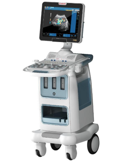
Esaote Biosound MyLab 40
The Esaote Biosound MyLab 40 ultrasound system is one of Biosound's powerful stationary ultrasound units. Specializing in Cardiovascular applications and images, this system is armed with advanced imaging technology, a myriad of ultrasound transducers, and can even be configured and updated to be a shared service and more versatile system.
When measuring an ultrasound system, two crucial factors include price and imaging technology. The MyLab 40 offers both extremely fair prices, and wildly advanced image enhancing technology to boot. 3D/4D (live 3D) images, TEI (tissue enhancement imaging), XView Speckle Reduction Imaging (SRI), XStrain (to enhance Cardiac images) are some foundational and integral technologies this system employs. Other crucial features include Tissue Velocity Mapping (TVM - to best display motion imaging), X4D Real Time 4D imaging, MView (similar to compound imaging) and integrated Stress Echo options.
The MyLab 40 is compatible with scores of ultrasound probes and transducers - over thirty of them! Many of these transducers are compatible with some other extremely popular Esaote Biosound ultrasound systems. This makes life considerably more economical, simple and straightforward for diagnosticians who wish to rotate between Esaote Biosound ultrasound systems. Instead of having to purchase new probes for each system, simply disconnect and reconnect with whichever system you wish to employ.
There are a number of types of probes that the MyLab 40 employs, ranging from endocavitary to linear array probes, phased array to curved array transducers. It also utilizes specialized Esaote probes that are more versatile and advanced with higher frequency ranges than other probes known as AppleProbes.
Ultrasound Features and Specificities:
- 17" and 19" TFT-LCD monitors available
- 200-degree endovaginal transducers
- Shared Services
- Appleprobe ergonomic transducers
- TEI Tissue Enhancement Imaging
- CMM Compass M-Mode
- XView speckle reduction imaging
- Advanced CnTI contrast imaging
- XStrain strain-rate imaging for myocardial analysis
- TVM Tissue Velocity Mapping for LV motion analysis
- AutoEF Automatic Ejection Fraction calculation
- Integrated Stress Echo
- MView combined standard and steered ultrasound imaging to detect all structures
- Tp-View trapezoidal imaging expanded view with linear transducers
- VPan panoramic extended field of view
- RF-QIMT Quality Intima Media Thickness
- X4D Real-Time 4D for advanced OB/GYN volumetric scanning
- DICOM
- Integrated CD/RW and USB port for exporting images and clinical data
- NT Nuchal Translucency/Nuchal Thickness calipers
Compatible Ultrasound Probes Transducers:
- MyLab CA631 Curved Array Probe
- MyLab CA541 Curved Array AppleProbe
- MyLab CA431 Curved Array Probe
- MyLab CA430 Curved Array Probe
- MyLab CA1421 Curved Array Probe
- MyLab CA123 Curved Array Probe
- MyLab C5-2 R13 Curved Array Probe
- MyLab BC431 Curved Array Probe
- MyLab BL433 Linear Array AppleProbe
- MyLab LA332 Linear Array AppleProbe
- MyLab LA435 Linear Array Probe
- MyLab PA240 Phased Array Probe
- MyLab PA121 Phased Array Probe
- MyLab BE1123 Endocavitary Probe
- MyLab EC1123 Endocavitary Probe
- MyLab IOE323 Hockey Stick Intraoperative Probe
- MyLab TEE132 TEE Probe
- MyLab Pencil CW2 Non Imaging Pencil Probe
***For a comprehensive list of all the compatible transducers from MyLab, click HERE**
F.A.Q.
Great question! The Esaote Biosound MyLab 40, before being updated or upgraded is a cardio-vascular ultrasound machine. Once upgraded, however, the machine becomes a shared service system.
Absolutely - especially if you're going to upgrade the system. When you do there's a variety of applications that can be projected in 3D/4D
- The MyLab 40 is compatible with the MyLab BL433 Linear Array AppleProbe, MyLab LA332 Linear Array AppleProbe and the MyLab CA541 Curved Array AppleProbe
XView/ Speckle Reduction Imaging works by changing the frequency of the sound into different beams (as opposed to one stronger blast), lowers the speckle that echoes back to the image, for the larger the beam, the more speckle. Another method includes lowering the beam’s power entirely – sacrificing slight image resolution but also preventing more speckle damage from perverting the image.