
What is Doppler Imaging?
Ultrasound doppler imaging is the ability to estimate and measure blood flow through various veins, arteries and vessels. Generally portrayed as a moving picture on an ultrasound system screen, one can usually recognize a doppler test from the color flow that is visible on the ultrasound image. The color in the image can be interpreted based on measuring blood movement in the specific area being photographed.
Doppler imaging is different than conventional ultrasound imaging in one fundamental way: It doesn’t actually image any structures. Conventional ultrasounds provide images for various structures, organs and veins to diagnose growths, breaks, structural problems, and many other potential ailments. Doppler imaging, on the other hand, projects images simply of blood flow.
Due to the non-invasive and non- radioactive nature of the ultrasound doppler imaging, it is a methodology that is globally established and revered. Instead of utilizing radiation or invasive features, the doppler functions the same way as other ultrasound imaging equipment does; employing high pitched sound waves that reflect and are translated into colors, images and various movements.
Doppler Ultrasound Imaging
Doppler Imaging’s Service:
Doppler imaging is different than conventional ultrasound imaging in one fundamental way: It doesn’t actually image any structures. Conventional ultrasounds provide images for various structures, organs and veins to diagnose growths, breaks, structural problems, and many other potential ailments.
Doppler imaging, on the other hand, is applied to detect blood flow and various potential hazards that can occur within the veins, arteries and vessels. Doppler imaging is generally used to detect blood clots, identify poorly functioning valves in veins, determine whether an artery is blocked, or recognize decreased blood circulation throughout the body. All of these potential threats to one’s health and life can be observed and prevented through use of doppler imaging.
There are different applications that people employ doppler imaging for: cardiac doppler, for example, and examining the blood flow to and from the heart is popular and extremely crucial part of cardiological examinations.
Other popular doppler applications include transcranial doppler (which tracks blood flow through the brain and the head), vascular, and general veins and arterial doppler.
Understanding Different Doppler Imaging Modes:
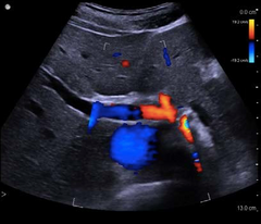
Once upon a time, before modern ultrasound technology, it was considered advanced for an ultrasound system to have color doppler imaging. Color doppler has two colors, red and blue. Using these colors, it became easier to perform exams and diagnose ailments accurately. Some older ultrasound systems, however, don’t have color doppler, and rely on a black and white screen to diagnose patients. Nowadays, thankfully, nearly all ultrasound machines uses color doppler.
In order to understand how doppler imaging works, one must understand how to identify what the colors on the ultrasound system indicate. The colors on the screen represent blood flow that is in motion. Blood moving towards the transducer is red, and blood moving away is blue. The lighter the shade of red or blue, the more rapid and quick the blood flow. The darker the shade, however, the slower the blood is moving.
Power doppler, a newer and more advanced form of doppler imaging, is used to provide doppler images for areas that are more difficult for conventional doppler modes have a difficult time capturing. It is used for small vessels, kidneys, and the brain. Although it has become more common in newer ultrasound systems, not all ultrasound machines employ power doppler options.
Doppler Transducers:
Every ultrasound manufacturer makes their own doppler probes, generally named “CW” (standing for continuous wave, a description of doppler images), followed by the frequency range. For example, the Mindray CW2s – which is short for Mindray continuous wave 2 MHz. probe. Most doppler probes, regardless of the manufacturer, are given names that are all similar to this example.
Doppler transducers have several other nicknames that individuals in the medical world refer to them as. Some of these nicknames include “blind probes”, “pedoff probes”, “continuous wave probes”, and obviously “doppler probes”. Any of these names are common among the medical community, and are used interchangeably.
Many states and insurance companies within the continental United States require that a diagnostician or sonographer that is purchasing an ultrasound machine must include a doppler continuous wave transducer with the machine. This requirement further emphasizes the importance of employing doppler imaging.
A.M.E. Ultrasounds offers a variety of ultrasound machines that are capable of providing color doppler imaging including the Medison Accuvix A30, the Philips HD9, and the GE Logiq V2 portableultrasound system.
There is always the concern, particularly when it comes to medical equipment, regarding the safety of the devices and the potential side effects that occur as a result of employing these machines.
It is important to know that ultrasound machines are known to be completely harmless. They function through the utilization of sound waves emitting from the probe or transducer. These sound waves are then translated into images by the ultrasound machine or system. There is no need to fear when using ultrasound machines – it is a quick, easy and efficient way to diagnose and help determine a patient’s ails.
At A.M.E. Ultrasounds we pride ourselves on providing the highest quality customer service. If there is a particular topic you would like to read about, learn more about, or would be interested in, please feel free to contact us!
If you’d like to learn about the author, click HERE
If you’d like to contact us, click HERE
Sincerely,
Your team at A.M.E. Ultrasounds
Ephraim@ameultrasounds.com


