A-Mode, B-Mode and C-Mode:
These two modes are simple in nature. A-Mode creates a display that graphs discoveries in the optic nerves. Using an X (depth) and Y (amplitude) axis, A-Mode drafts a one-dimensional presentation that obtains it’s information from the sound waves in the direction that the probe is facing.
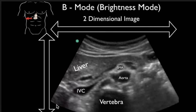
B-Mode, also known as 2D mode, presents a two dimensional demonstration. The brighter the image, the more intense and focused the echo (which is the reverberation of sound waves that the transducer emits) is. Like nearly all other ultrasound images, the position of the image is contingent on the angle that the transducer is placed.
C-Mode functions similarly to B-Mode, although it has not been been as developed to full potential. Using data and a range of depth from A-Mode, the transducer then moves to B-Mode (or 2D mode) and examines the whole region at the depth originally employed in two dimensional imagery.

B – Mode
M-Mode:
M-Mode functions based on time – similar to a stop-motion video. Rapidly emitting pulses, the ultrasound takes either A-Mode images or B-Mode images, making the expansive collection of pulses similar to an ultrasound video. This technique is often utilized to regulate the velocity of organ structures. M-Mode serves a different purpose than A-Mode, B-Mode and C-Mode by employing a series of pictures and observing the difference over a span of time.
Doppler Mode:
Although we have already elaborated on doppler imaging in our previous blog “How it Works: Doppler Ultrasound Imaging”, we can quickly recap it’s functionality. Applying ultrasound technology, diagnosticians are able to estimate and measure blood flow through various veins arteries and vessels. Recognizable by the moving colored images, red blood flowing towards the transducer and blue flowing away, the darker color slow moving blood and lighter color quick moving blood.
At A.M.E. Ultrasounds we pride ourselves on providing the highest quality customer service. If there is a particular topic you would like to read about, learn more about, or would be interested in, please feel free to contact us!
If you’d like to learn about the author, click HERE
If you’d like to contact us, click HERE
Sincerely,
Your team at A.M.E. Ultrasounds
Ephraim@ameultrasounds.com