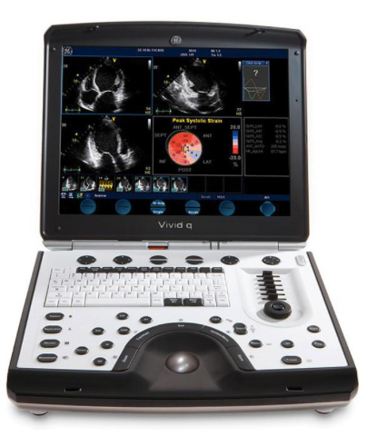
A new advance in portable echocardiography, extends the reach of medical professional to diagnose cardiovascular anatomy and LV function by adding more quantitave tools and pushing the boundaries to the highest levels of image quality. The Vivid q provides great accuracy and diagnostic confidence - while increasing your productivity. As part of the GE Vivid ultrasound series:
- The addition of the M4S-RS matrix array transducer produces enhanced 2D images for adult cardiac applications.
- Coded Octave imaging takes GE's image quality to the next level, enhancing your images for color flow and Doppler imaging.
- Smart Depth automatically adapts imaging parameters to help newer users see better results, expert operators save time and increase standardization among users.
- Smart Stress improves workflow, shortens optimization time and provides reproducibility for review, wall-segment scoring and reporting.
Vivid q builds on the many industry leading features and technologies of Vivid i with more performance, new quantitative analysis tools and a wider range of applications.
- Provides the most commonly used parameter to describe the LV function, the Ejection Fraction (EF).
- Assists in finding the endocardial border.
- Reduces the dependency of "seeing" the border in each image by analyzing and tracking the myocardial tissue.
- Automatically locates the end systolic and end diastolic frames.
Automated Function Imaging (AFI)
- Supports LV function analysis, visualizing the longitudinal wall shortening and lengthening and highlighting a segment's contraction.
- Can potentially be used to differentiate disease from non disease segments.
- Decreases LV function assessment variability, provides clinical decision support and streamlines workflow.
Tissue Velocity Imaging (TVI) and Tissue Tracking (TT)
Show tissue velocity and displacement in the direction of the ultrasound beam with high temporal resolution, and also visualize short events.Tissue Synchronization Imaging (TSI)
Translates comprehensive quantification into an easy-to understand image demonstrating mechanical synchronicity of different myocardial segments.Intracardiac Echo (ICE) imaging
Combining exceptional detail and quality of information with advanced interventional echocardiography ultrasound ICE imaging technology, Vivid q opens up a new window to the heart.- ICE catheters deliver excellent image quality and real-time visualization of cardiac structural anatomy, with therapy catheters for monitoring and guidance during interventional procedures.
- ICE gives you a better understanding of structural orientation during trans-septal puncture procedures to help you avoid clinical complications.
Shared Services
Vivid q's compact, laptop size and light weight make it easy to take exceptional ultrasound imaging performance wherever it's needed.- Conduct more vascular and abdominal exams with Vivid q's comprehensive set of linear and convex transducers.
- Display blood flow with 2D-like spatial resolution and no color-flow-imaging artifacts with B-Flow and Blood Flow Imaging (BFI).
- Measure the carotid artery's intima-media thickness quickly and accurately for early information on atherosclerosis risk with the IMT analysis package.
- Use Wide Aperture to improve the signal-to-noise ratio and spatial resolution for better penetration in deeper structures.
OR/Anesthesia
- Address perioperative needs with transthoracic examinations under challengIng conditions.
- Enable monitoring with the help of adult or pediatric TEE.
- Support saphenous vein harvesting and carotid evaluations.
- Use the intra-operative probe to support specific diagnoses in the OR during and directly after open-heart surgery.
- Share images remotely on any PC with the eVue option for efficient, convenient consultations.
Pediatric Echocardiography
- Examine children of all ages, including newborns, without compromise.
- Choose from a wide range of sector, micro convex, linear and transesophageal transducers plus a specific ECG cable.

